Title
Kurtis D. Borne
|
Supervisors: |
Artem Rudenko: Assistant Professor of Physics |
|
|
|
Daniel Rolles: Assistant Professor of Physics |
|
Kansas State University Physics Department REU Program

This program is funded by the National Science Foundation through grant number PHYS-1461251. Any opinions, findings, and conclusions or recommendations expressed in this material are those of the author(s) and do not necessarily reflect the views of the National Science Foundation.
Project Overview: Studying
nanoscale properties of molecular motion, energy, and geometry has developed
hand in hand with laser optics. Evidence of this is best conveyed with the
excitement over the “femtosecond pump-probe” experiments that are being
performed here in the James R. Macdonald Laboratory at Kansas State University.
Under the direction of Artem Rudenko, Daniel Rolles, and many others I studied
the fundamentals of single pulse laser-molecule interactions with
various important organic compounds. We observed fragmentation patterns and
energies associated with such interactions. Inevitably,
the results we found from this single pulse
procedure will be applied to more sophisticated pump-probe experiments. We will be able to
perform these experiments with more efficiency and accuracy than ever before
with the help from a newly equipped interferometric delay stage, which I helped
build.
1. Background and Motivation:
A
great deal of effort has been invested into the study of light induced
isomerization of various organic structures. Understanding the molecular
dynamics of such ultrafast and tortuous phenomenon will convey the underlying
mechanisms for biological properties such as isomerization of receptor
molecules in the eyes of animals or the photosystems of plants (1),(2). Because of this budding
interest, optical and molecular physicists have experimented with the fairly
simple organic molecule 1,3-cyclohexadiene ![]() . Over
five decades have been spent studying this molecule, and nearly one hundred
papers have been published with many enlightening results about this molecule.
High time-resolve experiments have revealed the necessary conditions for the molecules
signature rotational motion,and
the extremely quick transition times to higher order molecular energy levels
have been measured. The desire to study this molecule still pervades because
higher order energy levels still remain unexplored and the carbon-ring nature
of Cyclohexadiene acts
as a stepping stone for more complicated organic molecules (3). Also, the
ring-opening isomerization can play a crucial role in developing molecular
switches for whole new levels of technology (4).
. Over
five decades have been spent studying this molecule, and nearly one hundred
papers have been published with many enlightening results about this molecule.
High time-resolve experiments have revealed the necessary conditions for the molecules
signature rotational motion,and
the extremely quick transition times to higher order molecular energy levels
have been measured. The desire to study this molecule still pervades because
higher order energy levels still remain unexplored and the carbon-ring nature
of Cyclohexadiene acts
as a stepping stone for more complicated organic molecules (3). Also, the
ring-opening isomerization can play a crucial role in developing molecular
switches for whole new levels of technology (4).
But
you have to learn to walk before you can run. So, instead of implementing the
mentioned high time-resolve methods of pump probe microscopy, we
ionize the molecule using a single pulse 500 nm laser. A net absorption of one
or two photons from the laser pulse can cause ionization to occur. After this
ionization, what results is positive charge distributions throughout the
geometry of the molecule. These newly spawned positive ions will of course
repel each other in an ultrafast dynamic called Coulomb Explosion (5). For my overall goal, I wish to observe
the types of resulting fragments and associated kinetic energy released after Cyclohexadiene goes through this Coulomb Explosion.
There are various methods in order to achieve this goal, mostly involving time of flight mass –to-charge spectroscopy. Also we
will use PIPICO and TRIPICO
analysis, with which one can identify the two-body and three-body
fragmentation, respectively, of the parent
cyclohexadiene molecule after Coulomb Explosion.
If all the fragmentation species can be identified, and the results adhere to
the known parameters of the experiment, matching the previous results of
pathway isomerization, I can consider this experiment a success.
2. Setup and Experimental Technique:
2.1 PULSAR
For
ionization of molecules, high intense light sources are necessary. In order to
probe or control intermolecular interactions, ultrafast light sources are
necessary. Fortunately JRM labs is equipped with the state of the art PULSAR Laser
for such purposes (Pulsar, standing for Prarie
Ultrafast Light Source for Attosecond Research). For this experiment, we focused the laser beam
onto a parabolic mirror in order to focus it to the accuracy of a few microns.
This assures that laser-sample interaction has high probability. It also helps
reduce the occurrence of multi-molecule ionization. We desire only a single
molecule to interact with each laser pulse, in order to prevent false
coincidences, where it appears that two fragments originate from the same
parent molecule, when they actually don’t.
2.2 COLTRIMS
In
order to observe fragmentation patterns after laser interaction, we utilized
COLTRIMS techniques (COLTRIMS stands for COld Target
Recoil Ion Momentum Spectroscopy). The COLTRIMS system begins with the ejection
of a supersonic gas jet, which is so cold that the thermal momentum spread of
the molecules is essentially negligible (5). The necessity of such cold
temperatures is so that we can utilize the conservation of momentum, described
later.
The
COLTRIMS setup also contain an ultrahigh vacuum chamber, that uses a five stage
differential pumping mechanism to assure that residual and unwanted particles are
removed. After pumping, the chamber pressure is about ![]() Torr, or about
Torr, or about ![]() atmospheric units. This is absolutely necessary; we only want
detection 1-3 cyclohexadiene ionization, and data
reading would be nearly impossible if a lot ambient molecules were also
ionized.
atmospheric units. This is absolutely necessary; we only want
detection 1-3 cyclohexadiene ionization, and data
reading would be nearly impossible if a lot ambient molecules were also
ionized.
Within
the vacuum chamber, an adjustable electrostatic field is applied. The field is
used to increase the kinetic energy of the ions, as well as guide them onto a
detector at the end of the chamber. The electric field is generated by uniformly
varying the voltage between the anode and the cathode detectors.
Ionization
can be considered instantaneous, so at the moment of laser triggering, time
values begin to be calculated. The moment of laser triggering is determined by
a photodiode before entering the vacuum chamber. The time is calculated from
the moment of laser incidence until the ions are detected on microchannel plate
(MCP). The highly energized ion collides with the detector. Then a large chain
reaction of highly energized electrons come flying off from the detector. This
chain reaction makes it easy to determine the “stop-time” for the data
acquisition.
At
the back of the MCP is a delay line detector, which determines where the ion
lands on the detector. The way it works
is taking advantage of time differences between detection. The avalanche of electrons on the MCP will
send out a signal in both directions to the edge of the detector. The
propagation speed of this signal is constant in all directions. If the impact
is off-center, the signal will be processed at different times, and the
difference between these times has a direct relationship to the position on the
detector.
![]() Using detection time-difference to calculate
the X-coordinate.
Using detection time-difference to calculate
the X-coordinate.
![]() Using detection time-difference to
calculate the Y-Coordinate.
Using detection time-difference to
calculate the Y-Coordinate.
All
the values we wish to know from the light-matter interaction can be determined
by momentum and energy of the fragments. In order to calculate such things, it
is necessary to calibrate the time-of-flight and determine what fragmentation species
result after coulomb explosion.
2.3 Calibration
In
order to calibrate the time-of-flight detected, it helps to determine some
theoretical values. This can be done easily by simply solving for the equation
of motion of any ion. We begin by noting that the only force the ion
experiences is that applied to the electric field ![]() .
.
![]()
![]()
![]()
Solving
the quadratic equation for ![]() gives us a value for time-of-flight…
gives us a value for time-of-flight…
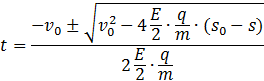
…
and plugging in the appropriate parameters where we
know that the initial position and velocity is zero…
The
above derivation makes the erroneous assumption of linear motion for the
fragment in the chamber. To adjust for this, we add an offset time ![]() to the above time of flight expression, which
is approximately the same for all ions.
Now we can calibrate the time-of-flight spectrum, here we look at two
high incident fragments and their distinctive time-of-flights, and solve for
the two constants in the equations below.
to the above time of flight expression, which
is approximately the same for all ions.
Now we can calibrate the time-of-flight spectrum, here we look at two
high incident fragments and their distinctive time-of-flights, and solve for
the two constants in the equations below.
![]()
![]()
As
an example, for run 209, I saw a very distinct pattern for water ![]() and for the singly
ionized parent cyclohexadine
and for the singly
ionized parent cyclohexadine ![]() . Using these
parameters I was able to solve for the unknown constants in the above equations
. Using these
parameters I was able to solve for the unknown constants in the above equations![]() .
.

Then,
solving for the mass charge ratio ![]() , and inserting
time-of-flight values of high incidence, we can see what kinds of molecules
result from fragmentation.
, and inserting
time-of-flight values of high incidence, we can see what kinds of molecules
result from fragmentation.
![]()
Finally,
with proper orientation of positioning on the detector and time-of-flights
calibrated, we can solve for the initial momentum vectors of the fragments
after coulomb explosion in all three directions.
![]() Momentum in the z-direction.
Momentum in the z-direction.
![]() Momentum in the
x-direction.
Momentum in the
x-direction.
![]() Momentum in the y-direction.
Momentum in the y-direction.
Calculating
the momentum of the fragments is important not only for the sake of knowing the
momentum and energy of fragments from coulomb explosion, it also helps us
distinguish good detection events versus bad ones. The “good events” are those
that fulfill the conservation of momentum requirement, specifically those
momentum vectors that sum to zero. This is justifiable for a few reasons; know
the initial momentum of the parent molecule is zero (see discussion of gas jet
above), and we know the momentum absorbed photons and emitted electrons will be
negligible in comparison to the ions after acceleration in the chamber.
3 Data
and Discussion:
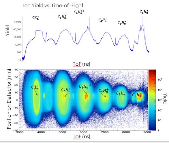
As
the Time-of-Flight spectrums above show, a lot of incidence has occurred in
this experiment. However, we are primarily interested in cases of the parent cyclohexadiene into two and three ionic fragments.
Observing these cases and adhering to the COLTRIMS measurement techniques, we
can look at released energy values, the geometry of breakup and compare them to
the predicted isomerization that is discussed in the theory section of this
report. The techniques of two-body
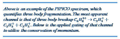
fragmentation is called PIPICO. The techniques of three body breakup are called
TriPICO. All these techniques are discussed here.
3.1 Two
Body Breakup:
When
the parent cyclohexadiene loses two electrons, the
resulting charge distribution could be in such a way that two positively
charged ions result. Because of their like charges, they will repel each other
with the energy determined by the Coulomb potential, converting such energy into
kinetic energy as they fly apart. This interaction is the aforementioned Coulomb explosion for two ions. Cases
for this two-body breakup can be seen on a PhotoIon-PhotoIon-Coincidence (PIPICO) spectrum.
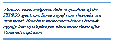
PIPICO spectrum
shows cases where two species result from the same laser shot. The x-axis
signifies the first ion that is detected, it will always have a smaller
time-of-flight. Therefore it will always be the smaller fragment. The y-axis
signifies the second ion that is detected, so it will always be the larger of
the two fragments. Cases of high incidence with the linear fashion shown on the
color gradient signify the two-body breakup that fulfills the conservation of momentum
conditions.
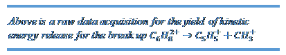
The
first annotated coordinate shows the events where the parent molecule is doubly
ionized and fragments as ![]() . The highest yield
for the kinetic energy released from this coulomb explosion centers around 3.00
eV to 3.20 eV, depending on the parameters of the laser and the applied
electric field.
. The highest yield
for the kinetic energy released from this coulomb explosion centers around 3.00
eV to 3.20 eV, depending on the parameters of the laser and the applied
electric field.
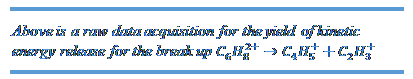
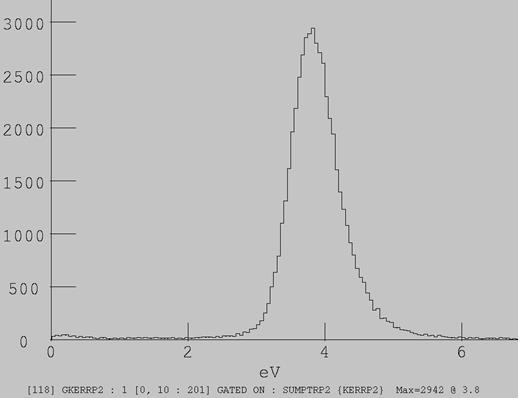
The
second annotation shows the breakup![]() . This has a slightly
larger kinetic enrgy associated with it, centering around 3.80 eV.
. This has a slightly
larger kinetic enrgy associated with it, centering around 3.80 eV.
3.2 Three
body breakup:
In
cases where the cyclohexadiene is triply ionized, a
coulomb repulsion between three fragments will occur. These cases have much
more interesting results because a lot of different geometries occur, even for
the same ionic resultants. Also, the types of fragments reveal possible
chain-opening isomerization that has been discovered in previous experiments,
as discussed in the theory section.
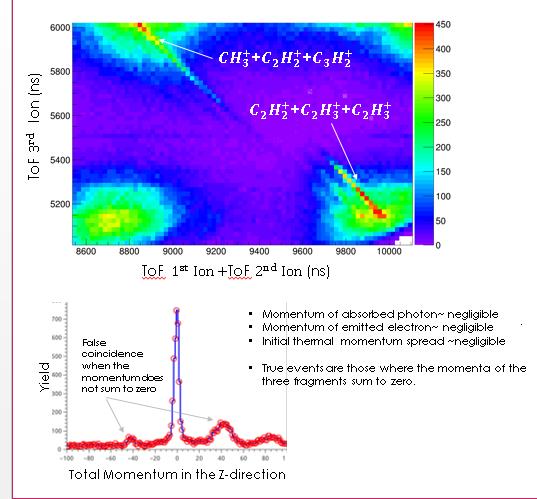
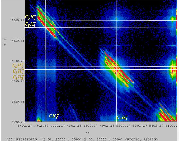
The
TriPICO spectrum shows cases where three species
result from a single laser incidence. The x-axis shows the time-of –flight for
the first ion that is detected. The y-axis is the sum of the time-of-flights
for the second and third ion that is detected. Once again, cases of high
incidence with the linear fashion shown on the color gradient signify breakups
that fulfill the conservation of momentum we implement.
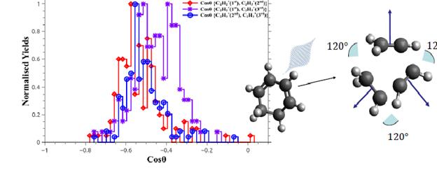
![]() The most apparent
result of this TriPICO analysis is the symmetric
breakup,
The most apparent
result of this TriPICO analysis is the symmetric
breakup, ![]() . In cases of this
symmetric breakup, we observe different distributions of the kinetic energy
amongst the fragments, as well as different geometries of the breakup. In order
to quantify this phenomenon 2D-rendered
. In cases of this
symmetric breakup, we observe different distributions of the kinetic energy
amongst the fragments, as well as different geometries of the breakup. In order
to quantify this phenomenon 2D-rendered
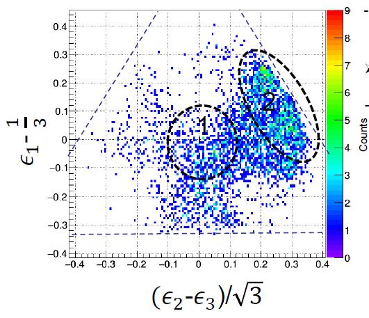
The
above diagram is a Dalitz Plot. Using this plot allows us to read
the energy distributions of three fragments on a single diagram. Areas of high
incidence determined by the color gradient show which fragment received more
kinetic energy after Coulomb explosion, depending on the events position on the
inlaying circle. A brief description on how Dalitz
Plots are formulated is in order:
0 0 G F E D![]()
![]()
![]()
![]()
![]()
![]()
![]()
![]()
![]()
![]()
![]()
![]()
![]()
![]()
![]()
![]()
![]()
![]()
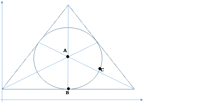
Dalitz Plots are defined in terms of a particles reduced energy. Reduced energy is a unit
less value determined by proportionality of momentums.
![]() Definition of reduced
energy
Definition of reduced
energy
![]() All reduced energies
of the fragments are normalized
All reduced energies
of the fragments are normalized
·
The
axes of the Dalitz plots are defined by differences
of the particles reduced energies.
![]() Horizontal axis of a Dalitz plot
Horizontal axis of a Dalitz plot
![]() Vertical axis on a Dalitz plot
Vertical axis on a Dalitz plot
In
our calibration procedure for symmetric breakup, we assigned fragment ![]() as
as ![]() and fragments 2 and 3 as
and fragments 2 and 3 as![]() .
.
The
Dalitz Plot presenting the data above shows two cases
of symmetric breakup.
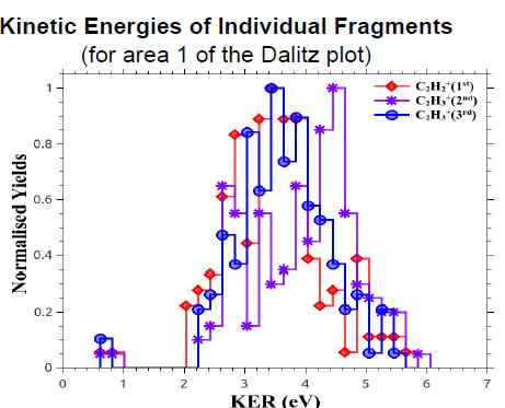
1. This is the case of
equal energy distribution and equiangular geometry. This is the scenario that
is expected for coulomb explosion of a closed ring. Plotting the energy
distribution for all three fragments, we see a value of about 3.8 eV. Also, the angular distribution of these
fragments has a peak of the obvious ![]() .
.
2. This scenario is the
case of procedural breakup. Where a ![]() ion is left stationary. This case gives hint
of possible ring opening of CHD to HT isomerization. This isomerization has
been witnessed before, and suppression of this kind of isomerization can be
done when a delayed laser pulse hits the molecule 50 fs after initial laser
excitation [6].
ion is left stationary. This case gives hint
of possible ring opening of CHD to HT isomerization. This isomerization has
been witnessed before, and suppression of this kind of isomerization can be
done when a delayed laser pulse hits the molecule 50 fs after initial laser
excitation [6].
4. Review
and Conclusion
In
this work, we used a 25 fs laser pulse in order to ionize the ring-structured
molecule 1,3-cyclohexadiene. Frequent cases of
multiple ionization occurred, which lead to two or three charged ions repelling
each other. This repulsion lead to intense dynamics associated with the release
of kinetic energy in a phenomenon called coulomb explosion.
We
saw frequent cases of two body break up: channels of ![]() and
and
![]() . Also, we focused on the rich information obtained from
symmetric three body breakup of
. Also, we focused on the rich information obtained from
symmetric three body breakup of ![]() . In this three-body
coincidence, we see a lot of cases of equiangular geometry and equal kinetic
energy among all three fragments. Other cases of unequal energy sharing give
hints of a pathway in which the molecular geometry varies during ionization.
. In this three-body
coincidence, we see a lot of cases of equiangular geometry and equal kinetic
energy among all three fragments. Other cases of unequal energy sharing give
hints of a pathway in which the molecular geometry varies during ionization.
So now, we have a few fragmentation patterns
that we would like to observe further. With the new delay stage attached and ![]() harmonic generator, we will be able to perform
pump-probe laser microscopy. Using this technique, a single infrared pulse will
induce excitation of the molecule to higher energy levels, and after a certain,
controllable, delay time, a second x-ray will ionize it. Then we can perform
these Coulomb Explosion techniques to determine the time-dependent dynamics of
the molecule.
harmonic generator, we will be able to perform
pump-probe laser microscopy. Using this technique, a single infrared pulse will
induce excitation of the molecule to higher energy levels, and after a certain,
controllable, delay time, a second x-ray will ionize it. Then we can perform
these Coulomb Explosion techniques to determine the time-dependent dynamics of
the molecule.
[1] Wang, Q., R. Schoenlein, L. Peteanu, R. Mathies, and C.
Shank. "Vibrationally Coherent Photochemistry in the Femtosecond
Primary Event of Vision." Science 2665184 (1994): 422-24.
[2] Zubik, Monika, Rafał Luchowski, Wojciech Grudzinski, Małgorzata Gospodarek, Ignacy Gryczynski, Zygmunt Gryczynski, Jerzy W. Dobrucki,
and Wiesław I. Gruszecki.
"Light-induced Isomerization of the LHCII-bound Xanthophyll Neoxanthin: Possible Implications for Photoprotection
in Plants." Biochimica Et
Biophysica Acta (BBA) - Bioenergetics
1807.9 (2011): 1237-243.
[3] Pullen
S.H., Anderson N.A., Walker L.A., Sension
R.J. “The ultrafast photochemical ring-opening reaction of the
1,3-cyclohexadiene in cyclohexane.” J. Chem. Phys. (1995) 108:556–63
[4] Geppert, D., L.
Seyfarth, and R. De Vivie-Riedle.
"Laser Control Schemes for Molecular Switches." Applied Physics B
Appl. Phys. B 79.8 (2004): 987-92.
[5] Maharjan,
Chakra Man. Momentum Imaging Studies of Electron and Ion Dynamics in a Strong
Laser Field. PhD thesis. 2007
[6] Ergler, Th., A. A. Rudenko, B. Feuerstein, K. Zrost, C.
D. Schröter, R. Moshammer, and J. Ullrich. "Fragmentation
of Molecules Studied with Laser-induced Coulomb Explosion Imaging and
Femtosecond Pump-probe Experiments." (2006): n. pag. Web.
Thanks: Thanks to professor Artem Rudenko and Daniel Rolles for all their direction in the research at every step of the way. And thanks to everyone in the group: Farzaneh, Balram, and Shashank. They were with me through all experimental procedurals. Without them I wouldn’t have even known how to upload data... Utuq, Seyyed, Xiang ( I miss you…), Yubaraj (wish I could have met you), and Jeff.
About Me: I attend College University of Nebraska of Omaha. I study Physics with a minor in Mathematics, and German. I like to sit on couches and listen to the Grateful Dead a lot…